
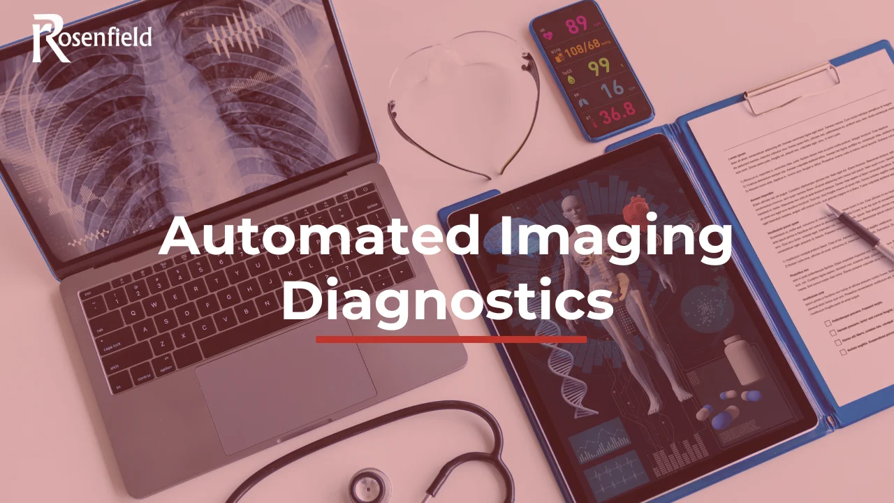
Automated Imaging Diagnostics
Medical imaging is witnessing an exceptional transformation due to the immense leap occurring in artificial intelligence technologies. Through smart systems, it has become possible to analyse visual data with accuracy and speed, whereas reliance was previously solely on human expertise. Automated imaging diagnostics has led to raising the level of diagnostic accuracy, the possibility of detecting diseases early, and also increasing speed diagnostics medical imaging, thus making healthcare more effective and inclusive.
What are automated imaging diagnostics?
Automated imaging diagnostics is the employment of AI algorithms, particularly deep learning technologies, to execute some tasks that were exclusive to human expertise, such as recognising patterns, how to make decisions, and analysing the minutest details within medical images.
These tools do not represent mere aids but constitute a system through which continuous learning is possible thanks to huge amounts of data, thus gaining accuracy and proficiency over time.
The goal of Automated Imaging Diagnostics is not to use the machine instead of the radiologist, but rather to perform tasks more efficiently and accurately through a radiologist augmented by AI to accomplish work and reduce the probability of clinical errors.
How AI powers diagnostic imaging today
Understanding How AI is Used in Radiology? in the UK begins with deep learning, a branch of machine learning that uses multi-layered neural networks, especially Convolutional Neural Networks (CNNs), capable of extracting complex patterns from images automatically. These algorithms are trained on thousands or millions of previously classified medical images, consequently having the ability to differentiate between healthy and diseased tissues with great accuracy and also identify the minutest lesions that the human eye might miss. Among the core applications of these technologies:
- Classification and Segmentation: Through which the type of lesion can be identified and its boundaries known with great accuracy.
- Feature Extraction: Called radiomics, it can detect the minutest patterns that cannot be seen with the naked eye.
- Disease Trajectory Prediction: Through which data is analysed to predict treatment response and clinical developments.
Benefits: speed, accuracy, early detection
There are multiple tangible benefits resulting from integrating AI into radiological practices, including:
- Accelerating work and increasing efficiency: Images can be interpreted in a shorter time ranging from 30% to 75%, and consequently reports can be written faster by a percentage starting from 30% to 50%.
- Raising diagnostic accuracy: Algorithms help reduce errors resulting from work pressure or fatigue. Algorithms have demonstrated the accuracy of some models exceeding 94% for lesion segmentation and sensitivity reaching up to 95% for detecting pulmonary nodules.
- Enhancing early detection: Smart systems help monitor the first signs of disease such as cancer, thus potentially raising treatment chances significantly.
Use cases: emergency, pathology, ultrasound
The applications of AI in radiology are more comprehensive for many sub-specialties, including:
- Emergency cases: Including immediate analysis of brain scans to identify pulmonary embolism or detect strokes within a limited time, thus making strict decisions in a few minutes.
- Oncology: It has become possible to detect liver lesions and screen for breast cancer accurately, and also contribute to radiotherapy planning; therefore, it is considered one of the most mature applications.
- Pathology: Applications have contributed to analysing histological slides that help determine the tumour severity grade and classify it.
- Cardiovascular medicine: Algorithms are used in assessing coronary artery narrowing and also left ventricle segmentation, thus enhancing the accuracy of cardiac assessments.
Challenges: misdiagnosis risk, data bias, regulatory approval
Despite the development of AI in medical imaging and its immense capabilities, it faces some pivotal challenges, including:
Misdiagnosis and transparency
Models might carry errors for unfamiliar cases. In addition, the “black box” model hinders radiologists from understanding decision-making mechanisms. Therefore, explainable AI plays a crucial role in disease diagnosis by enhancing trust and transparency.
Data bias and poor generalisation
Algorithms might potentially fail when applied to different population groups if training data is not diverse or inclusive of all population categories, thus potentially exacerbating health disparities.
Regulatory hurdles
AI tools are subject to regulatory oversight by the FDA to guarantee safety and efficacy. These approvals can take a long and costly time, leading to slowing down the introduction of innovations to the market.
The Challenge of Data Preparation: The Essential Fuel for AI
One of the most prominent challenges in building AI models is data preparation. All identifying information must be removed from medical images before they enter the training phase to protect patient privacy and comply with global regulations.
Given the vast amounts of data that medical institutions handle—which can reach thousands of studies—manual anonymisation is exhausting, impractical, and prone to diagnostic errors in radiology.
To address this, advanced solutions like BriX provide an effective mechanism to anonymise data quickly and accurately, reducing the time required from weeks to just a few minutes.
Implementation steps for Trusts & radiology departments
The process of adopting AI is not just a new programme to be acquired, but rather an integrated strategic journey. To guarantee its success and reach the highest actual value, focus must be placed on some basic steps including:
Building a solid data governance framework
Clear policies must be set through which data can be handled while guaranteeing quality and protecting privacy.
Enhancing multidisciplinary collaboration
Close cooperation must be achieved between radiologists, IT engineers, and data scientists to be able to implement successfully and guarantee aligning solutions with clinical needs.
Algorithm validation
Internal or external validation must be conducted to ensure the actual performance of algorithms and guarantee suitability and accuracy, not relying solely on supplier claims.
Integrating solutions into existing workflow
Priority must be given to solutions compatible with PACS that can present results in a way that does not affect the radiologist’s concentration.
Training and adapting the workforce
It is preferable to provide appropriate training for both staff and radiologists to understand the limits of AI technologies and capabilities, thus enhancing their use as supportive tools, not replacements.
Conclusion
Automated imaging diagnostics are reshaping radiology by combining clinical expertise with AI-driven precision to improve diagnostic accuracy, speed, and patient outcomes. When implemented correctly, these technologies reduce error rates, optimise workflows, and support radiologists in delivering safer, more consistent care.
👉 Connect us at Rosenfield Health or book a demo to see how our AI-ready imaging and data solutions help NHS Trusts and radiology departments adopt automation securely, efficiently, and at scale.
FAQs
What does automated imaging diagnostics mean?
It is the employment of AI algorithms, specifically deep learning, which helps analyse medical images and detect diseases with great accuracy, knowing their classification and impact extent, and consequently working on making clinical decisions quickly after improving diagnostic quality.
Do automated diagnostics reduce diagnosis time?
Yes, significantly. Examination time can be reduced by up to 75% with the possibility of preparing reports quickly by 50% through identifying cases that have priority and medical imaging diagnostics automation tools.
Are automated diagnosis tools accurate?
Yes, automated diagnostics in radiology can achieve a lot of accuracy. Some studies have shown that trained algorithms can reach the highest levels of accuracy exceeding or surpassing human expertise, represented in breast cancer screening or detecting pulmonary nodules.